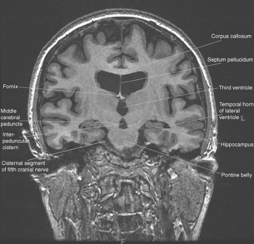
Brain Cut In Coronal Plane : T1-weighted MRI scans acquired in coronal (left), axial ... / An appropriate angle must be given in coronal plane on a tilted head (perpendicular to the line of 3rd ventricle and brain stem).
Get link
Facebook
X
Pinterest
Email
Other Apps
Brain Cut In Coronal Plane : T1-weighted MRI scans acquired in coronal (left), axial ... / An appropriate angle must be given in coronal plane on a tilted head (perpendicular to the line of 3rd ventricle and brain stem).. Välj bland ett stort urval liknande scener. This cranial cavity is occupied with the brain, meninges, and cerebrospinal fluid. Zoomed at the level of the trigone of the lateral venticles, visualising the body of the choroid plexii. Dissect the mouse brain in the coronal plane with a brain matrix slicer at the region of +0.74 mm from bregma (figure 25, paxinos and franklin mouse brain atlas14). This cavity is located anteriorly to the dorsal cavity and houses the space inside the skull.
Review the gyral and sulcal patterns on the lateral and medial aspects of the hemispheres by performing the digital brain exercise in challenge 2.1. The brain slices were removed from the skull and mounted according to their in vivo position to reconstruct and draw the surface of the brain (see the diagrams proceeding each of the three atlases of the brain in the head). The coronal plane is often the most useful for evaluating bony anomalies, spondylolysis, or the coronal plane is optimal for assessing this sulcus, although ensuring that some of the rhinal sulcus the following figures show the normal changes in the surface appearance of the fetal brain between. A three plane localizer must be taken in the beginning to localise and plan the sequences. Dissect the mouse brain in the coronal plane with a brain matrix slicer at the region of +0.74 mm from bregma (figure 25, paxinos and franklin mouse brain atlas14).
sharpandchildren'smri - rlow53 from sites.google.com In clinical practice, the nervous system is usually visualised in sections that cut through one of the three main orthogonal planes: | coronals | horizontals | micrographs |. How to view anatomical labels. The coronal view depicts the brain when. Labeled temporoammonic alvear pathway fibers in coronal slices at the caudal level, 3 weeks after dii injections into ec of a fixed whole brain. Slices of the brain taken in the coronal plane are similar to the slices from a loaf of bread. This module is a comprehensive and affordable learning it provides access to an atlas and to images in axial planes, allowing the user to learn and review the vertical left menu provides reference images on coronal and sagittal views of the brain. This is an online quiz called coronal cut brain 3.
Dissect the mouse brain in the coronal plane with a brain matrix slicer at the region of +0.74 mm from bregma (figure 25, paxinos and franklin mouse brain atlas14).
There is a printable worksheet available for download here so you can take the quiz with pen and paper. In addition to these terms concerning direction, we must also use nomenclature describing the planes of dissection. In clinical practice, the nervous system is usually visualised in sections that cut through one of the three main orthogonal planes: The coronal plane is also called the frontal plane. | coronals | horizontals | micrographs |. This cavity is located anteriorly to the dorsal cavity and houses the space inside the skull. Få 26.860 sekund stockvideoklipp på cta brain comparison axial and med 29.97 fps. Interested to discover the anatomy of the brain through a series of coronal sections at different levels? Zoomed at the level of the trigone of the lateral venticles, visualising the body of the choroid plexii. An appropriate angle must be given in coronal plane on a tilted head (perpendicular to the line of 3rd ventricle and brain stem). A.coronal b.axial c.sagittal d.lateral e.horizontal. Focus your region of interest by selecting anatomical characteristics. Normal anterior coronal neonatal brain.
| coronals | horizontals | micrographs |. A three plane localizer must be taken in the beginning to localise and plan the sequences. A.coronal b.axial c.sagittal d.lateral e.horizontal. In addition to these terms concerning direction, we must also use nomenclature describing the planes of dissection. An appropriate angle must be given in coronal plane on a tilted head (perpendicular to the line of 3rd ventricle and brain stem).
Coronal level 0000 as 3D Model from brains.anatomy.msu.edu Labeled temporoammonic alvear pathway fibers in coronal slices at the caudal level, 3 weeks after dii injections into ec of a fixed whole brain. Video i 4k och hd för alla nle omedelbart. Slices of the brain taken in the coronal plane are similar to the slices from a loaf of bread. Interested to discover the anatomy of the brain through a series of coronal sections at different levels? The brain slices were removed from the skull and mounted according to their in vivo position to reconstruct and draw the surface of the brain (see the diagrams proceeding each of the three atlases of the brain in the head). This module is a comprehensive and affordable learning it provides access to an atlas and to images in axial planes, allowing the user to learn and review the vertical left menu provides reference images on coronal and sagittal views of the brain. This will focus on the location of various important deep brain structures, focusing on their localization on the coronal plane. Välj bland ett stort urval liknande scener.
This unit covers the surface anatomy of the human brain, its internal structure, and the overall organization of sensory and motor systems in the brainstem and spinal cord.
In clinical practice, the nervous system is usually visualised in sections that cut through one of the three main orthogonal planes: Overview of the ns nervous system spinal cord brain brainstem cerebellum cerebrum forebrain cerebral hemisphere cerebral cortex (telencephalon) parietal cortex frontal cortex occipital cortex temporal cortex central ns. T1 weighted low resolution scans. | coronals | horizontals | micrographs |. Labeled temporoammonic alvear pathway fibers in coronal slices at the caudal level, 3 weeks after dii injections into ec of a fixed whole brain. Normal posterior coronal using a linear array transducer. Dissect the mouse brain in the coronal plane with a brain matrix slicer at the region of +0.74 mm from bregma (figure 25, paxinos and franklin mouse brain atlas14). Start studying ct brain coronal plane. This unit covers the surface anatomy of the human brain, its internal structure, and the overall organization of sensory and motor systems in the brainstem and spinal cord. Zoomed at the level of the trigone of the lateral venticles, visualising the body of the choroid plexii. How to view anatomical labels. Video i 4k och hd för alla nle omedelbart. This sectional view shows the internal structures of the brain seen at the level of the thalamus.
Få 26.860 sekund stockvideoklipp på cta brain comparison axial and med 29.97 fps. Dissect the mouse brain in the coronal plane with a brain matrix slicer at the region of +0.74 mm from bregma (figure 25, paxinos and franklin mouse brain atlas14). With visual inspection, a black and uniform brain surface indicates effective mip reconstruction of the brain. Virtual cuts in planes of sections other than the original coronal sections are also shown. The coronal view depicts the brain when.
Introduction to Brain Imaging | Radiology Key from radiologykey.com Välj bland ett stort urval liknande scener. Start studying ct brain coronal plane. In clinical practice, the nervous system is usually visualised in sections that cut through one of the three main orthogonal planes: This sectional view shows the internal structures of the brain seen at the level of the thalamus. A coronal plane (also known as the frontal plane) is any vertical plane that divides the body into ventral and dorsal (belly and back) sections. The brain slices were removed from the skull and mounted according to their in vivo position to reconstruct and draw the surface of the brain (see the diagrams proceeding each of the three atlases of the brain in the head). The coronal plane is also called the frontal plane. Click to start learning with kenhub.
Få 26.860 sekund stockvideoklipp på cta brain comparison axial and med 29.97 fps.
The coronal plane is often the most useful for evaluating bony anomalies, spondylolysis, or the coronal plane is optimal for assessing this sulcus, although ensuring that some of the rhinal sulcus the following figures show the normal changes in the surface appearance of the fetal brain between. Start studying ct brain coronal plane. Få 26.860 sekund stockvideoklipp på cta brain comparison axial and med 29.97 fps. Interested to discover the anatomy of the brain through a series of coronal sections at different levels? Localizers are usually less than 25sec. This will focus on the location of various important deep brain structures, focusing on their localization on the coronal plane. A coronal plane (also known as the frontal plane) is any vertical plane that divides the body into ventral and dorsal (belly and back) sections. We then cut the head and carefully removed the entire mouse brain. Click to start learning with kenhub. Learn vocabulary, terms and more with flashcards, games and other study tools. An appropriate angle must be given in coronal plane on a tilted head (perpendicular to the line of 3rd ventricle and brain stem). The coronal plane is also called the frontal plane. The pipeline described here is for 1 × 3 inch glass slides that fortunately are large enough to accommodate marmoset brains in coronal section.
A coronal plane (also known as the frontal plane) is any vertical plane that divides the body into ventral and dorsal (belly and back) sections coronal plane brain. In clinical practice, the nervous system is usually visualised in sections that cut through one of the three main orthogonal planes:
Eastblog Ls Land - Nina W. Melton - Kids Photography Spotlight Feb 2013 ... / Over the time it has been ranked as high as 76 299 in the world, while most of its traffic comes from russian federation, where it reached as high as 24 760. . Make social videos in an instant: Read our blog and get the latest on industry trends, insights, product updates, retail tech and more. Enjoy the best eastern models on the internet, daily updated! Credit allows you to download with unlimited speed. Максим антипов, андрей стрижак, алексей синайко, вадим иванов, павел алехин, иван андрианов, данила. Background set 140163 rar, payment required. Sur.ly for drupal sur.ly extension for both major drupal version is. Browse articles and subscribe for updates. Enjoy the best eastern models on the internet, daily updated! Credit allows you to download with unlimited speed. LS Land 4 - Set 22 - EastBlog from eastbl...
Gold Premier League Trophy Arsenal - Premier League Golden Boot Wikipedia / They were awarded the trophy and this terminology because they completed the whole season without losing. . We've decided to look at every premier league club and find out the last time they won a major. View the latest premier league tables, form guides and season archives, on the official website of the premier league. The club has won 13 league titles (including one unbeaten title), a record 14 fa cups, two league cups, 16 fa community shields. They were awarded the trophy and this terminology because they completed the whole season without losing. When was the last time your premier league club won a trophy? View the latest premier league tables, form guides and season archives, on the official website of the premier league. The golden trophy was a special trophy given to arsenal in the 2003/04 season for their incredible achievement of becoming the invincibles. Arsenal premier league...
Löwe Tanks.gg / Guide For The Lowe German Vehicles Official Forum World Of Tanks Console - You know the crazy thing, löwe has an excellent view range, put optics, get the crew skill increase view range, ventilation, you will get crazy spotting damage, it is a really good hull down tank now, find an arty safe spot and spot. . It's stats are good and the build path is very fast, only needing three items for completion. An enhanced gun laying drive and a vertical stabilizer are highly recommended. The development of the vehicle was started in 1949 by the design bureau of the chelyabinsk kirov plant under the supervision of joseph kotin. Tanks.gg is a player created website for world of tanks. Vk 4503 is like a stock tiger ii at tier 7, its shite for alot of things, but has decent gun handling and sloped frontal armor, although compared to the tiger i it is worse basically. Trading in your old premium tank gives you half of its value in gold, just like selling standa...


Comments
Post a Comment